The Inguinal Canal
The processus vaginalis, a peritoneal diverticulum, forms the basic structure of the inguinal canal, a tubular structure formed by multiple coverings of the anterior abdominal wall. It is an oblique passage approximately 4cm long extending inferiorly and medially through the anterior abdominal wall, superior to and parallel to the lower half of the inguinal ligament. It contains the ilioinguinal nerve together with spermatic cord in a male and the round ligament of the uterus in a female.
The inguinal canal is an oblique passage, which begins at the deep inguinal ring and continues to the superficial inguinal ring and is subsequently a region of weakness through which herniation can occur. However its physiologic mechanisms, including the oblique direction of the canal, aid in maintaining its integrity.
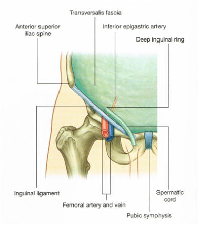 Figure 11. Deep inguinal ring and the transversalis fascia (Drake et al. 2010: p. 284).
Deep inguinal ring:
The deep inguinal ring is an oval opening, the beginning of the tubular evagination, located within the transversalis fascia, seen in Figure 11 (Drake et al. 2010), located halfway between the anterior superior iliac spine and the pubic symphysis and approximately 1.3cm superior to the inguinal ligament.
The spermatic fascia and the round ligament originate from this layer of transversalis fascia of the deep inguinal ring.
It is immediately lateral to the inferior epigastric vessels.
Superficial inguinal ring:
The superficial inguinal ring is a triangular-shaped defect in the aponeurosis of external oblique, seen in Figure 12 (Drake et al. 2010), with its base formed by the pubic crest and its apex pointing supero-laterally. Its medial and lateral margins are called crura with Its medial crus attaching to the pubic crest and the lateral crus attaching to the pubic tubercle. It forms the end of the inguinal canal and is located superior to the pubic tubercle. To strengthen and support it the medial and lateral borders are held together by intercrural fibres located towards its apex.
Scroll over both the labels and the structures in the image below to highlight them and/or reveal where they are located. By clicking on various structures descriptions will appear under some areas.
Figure 12. Superficial inguinal ring and the aponeurosis of the external oblique, adapted from (Drake et al. 2010: p. 285).
Borders of the Inguinal Canal:
The inguinal canal, seen in Figure 13 (Drake et al. 2010), has four boundaries associated with it; an anterior and posterior wall and a roof and floor. These boundaries are formed either by the fascia and muscles of the anterior abdominal wall or by the inguinal ligament.
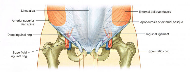
Figure 13. Inguinal canal (Drake et al. 2010: p. 284).
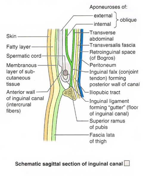 Figure 14. Inguinal canal and spermatic cord - Schematic sagittal section of inguinal canal (Moore et al. 2010: p. 206).
Anterior wall:
Formed by the aponeurosis of external oblique with a small component within the lateral third comprising internal oblique, seen in Figure 14 (Moore et al. 2010) which reinforces the canal laterally around the deep inguinal ring.
Posterior wall:
Formed by the lateral 2/3rds of the transversalis fascia and at the medial third by the conjoint tendon, which is formed by common insertion of transversus abdominis and internal oblique to the pubic crest and the pectineal line and reinforces the superficial inguinal ring.
Roof:
Fibres of internal oblique and the transversus abdominis arching over the contents of the inguinal canal
Floor:
The inguinal ligament formed from the inferior rolled edge of the aponeurosis of external oblique and medially the lacunar ligament, which reinforces the inguinal canal medially.
