Development of the Inguinal Canal
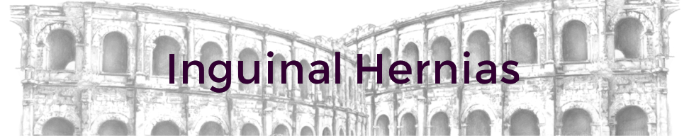
The inguinal canal is an oblique passage that extends inferiorly and medially through the anterior abdominal wall, superior to and parallel to the lower half of the inguinal ligament. It is created by an outpouching of the peritoneum, a peritoneal diverticulum described as the processus vaginalis. During development the gonads in both sexes must descend into the pelvic cavity from the superior lumbar region of the posterior abdominal wall where they originated, seen in Figure 5 (Moore et al. 2010). Mesenchymal cells condense to form the gubernaculum, an embryonic structure, which attach the caudal ends of the gonads, terminating in the inguinal region, between the internal and external abdominal oblique muscles, with labioscrotal swellings.

Figure 5. The testis attached to the posterior abdominal wall in a seventh week embryo (Moore et al. 2010: 205).
This tubular sheath projects through the peritoneal cavity between the anterior abdominal wall musculature, acquiring coverings from each and alongside the gubernaculum into the labioscrotal swellings. The pathway that the gubernaculum has formed guides the descent of the gonads where by in males the testis descend between the processes vaginalis, seen in Figure 6 (Moore et al. 2010) and the muscular layers of the anterior abdominal wall into the scrotal sac. The processus vaginalis normally regresses, with its distal part forming the tunica vaginalis, seen in Figure 7 (Moore et al. 2010), covering the surface of the testis and subsequently the connection and opening between the abdominal cavity and the processus vaginalis closes but failure to do so can result in an indirect (congenital) hernia in both males and females.
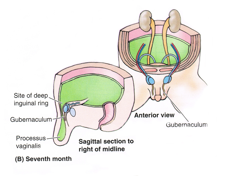
Figure 6. Processus vaginalis and testes passing through the inguinal canal in the seventh month (Moore et al. 2010: p. 205).
|
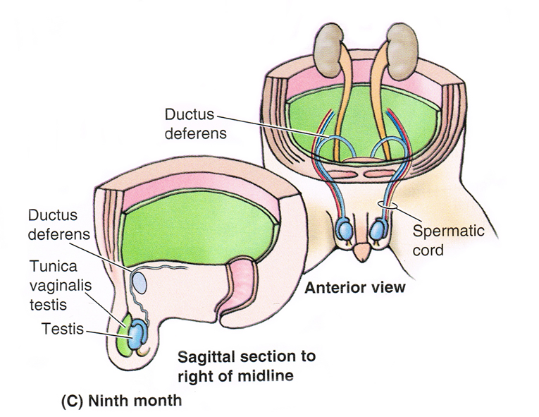
Figure 7. Obliteration of the stalk of the processes vaginalis has occurred in the ninth month (Moore et al. 2010: p. 205).
|
During male development a collection of structures pass along the inguinal canal to form the testis, which include the ductus deferens, vessels and nerves. These structures form the spermatic cord, which is surrounded by three concentric layers of fascia derived from the anterior abdominal wall; internal spermatic fascia, cremaster muscle and the external spermatic fascia respectively, which passes through the inguinal canal along with the ilioinguinal nerve.
However, in females the descent of the gonads is considerably less, where by the gubernaculum attaches to the primordial ovaries and the future labia majora, seen in Figure 8A (Moore et al. 2010). The ovaries finally lie just below the rim of the pelvis laterally within the pelvic cavity, with the gubernaculum subsequently forming the ovarian ligament and the round ligament, seen in Figure 8B (Moore et al. 2010), with the latter extending into the labia majora. Any protrusion of the peritoneum into the labia majora is termed the 'Canal of Nuck', which is analogous to the processes vaginalis in males.
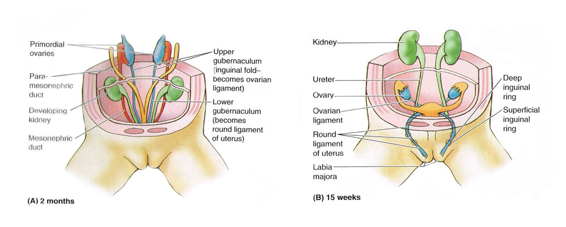
Figure 8. Formation of the inguinal canals in females. A. At 2 months the undifferentiated gonads (primordial ovaries) are located on the dorsal abdominal wall. B. At 15 weeks the ovaries have descended into the greater pelvis (Moore et al. 2010: p. 206).
In females the round ligament of the uterus, seen in Figure 9 (Moore et al. 2010) a remnant of the gubernaculum, the ilioinguinal nerve, the genital branch of the genitofemoral nerve and blood and lymphatic vessels pass through the inguinal canal.
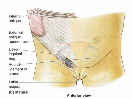
Figure 9. Formation of the inguinal canal in females. C. In the mature female, the processus vaginalis has degenerated but the round ligament persists and passes through the inguinal canal (Moore et al. 2010: p. 206).
Although the gubernaculum, seen in Figure 10 (Drake et al. 2010), is directly associated with the migration of the testes and the ovaries and subsequently with the migration of the testes through the inguinal canal, there is still controversy as to which role the gubernaculum plays in relation to the inguinal canal. Although the inguinal canal forms prior to testicular descent the gubernaculum does play a fundamental role in testicular migration.
Scroll over both the labels and the structures in the image below to highlight them and/or reveal where they are located. By clicking on various structures descriptions will appear under some areas.
Figure 10. Descent of the testes from week 7 (post fertilisation) to birth, adapted from (Drake et al. 2010: p. 283).
